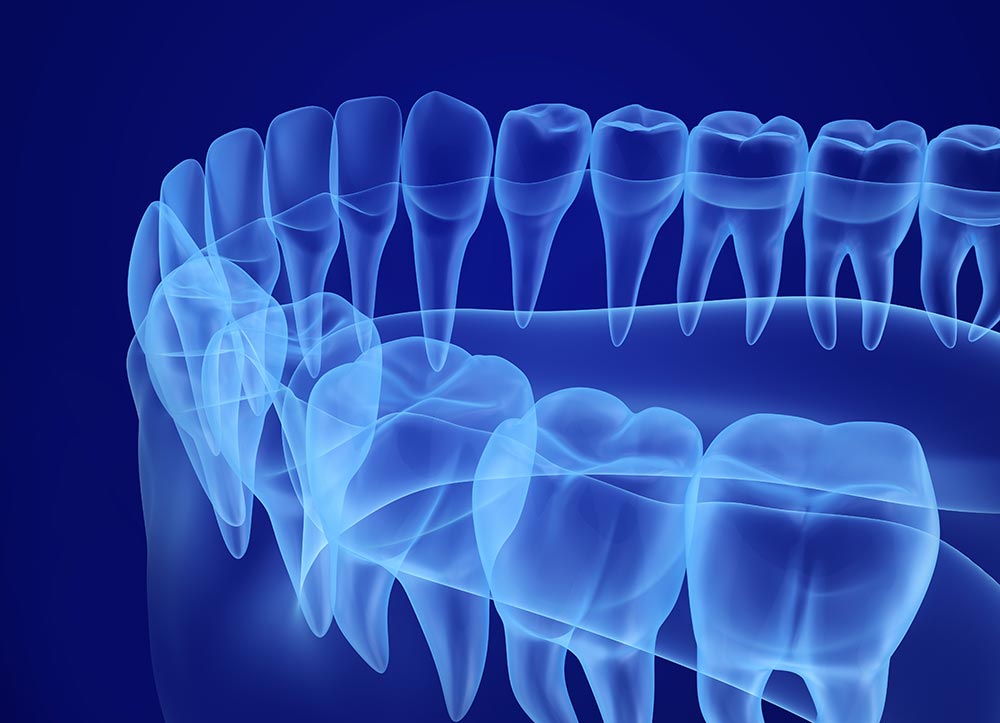
TRIDIMENSIONAL RADIOLOGY: WHAT IS IT
In various types of interventions, it is extremely important to resort to tridimensional radiology to examine the anatomical structures that will be involved. Radiological examinations in dentistry allow the dentist to make the correct diagnosis and thus choose the most suitable therapy to solve the patient’s problem.
In oral surgery and advanced implantology interventions, having an accurate image of the structures involved is useful as it allows planning and simulating the type of intervention that will be performed.
The medical staff of our dental clinic in Milan has noticed a clear improvement in the evaluation and resolution of the most common dental diseases through the use of this new technology. Furthermore, we also notice the satisfaction of our clients, who feel more relaxed and at ease in facing the analysis and treatment of their dental problems.
TRIDIMENSIONAL RADIOLOGY: CUTTING-EDGE DIAGNOSIS
Our dental clinic in Milan chooses high-level radiological equipment. We want to offer our customers maximum precision in diagnosis and explanation of the therapies and treatments proposed. This approach allows us to observe, from our patients, a more immediate and serene acceptance of the therapeutic path to be undertaken based on complete information.
In particular, we have introduced the use of instrumentation that uses one of the most renowned systems worldwide for tridimensional radiology. It is particularly practical for use in dental clinics. It allows obtaining radiological images such as orthopantomography (OPG or panoramic radiography) and CT scans performed with cone-beam technology.
This last tool allows the use of radiological studies with tridimensional reconstructions and guarantees excellent image quality. It also reduces patient exposure to X-rays.
MAIN DENTAL RADIOLOGICAL EXAMINATIONS
The main radiological examinations used by the dentist are:
- Endoral radiography, used to acquire the image of up to a maximum of three teeth on the same plate. It is used to confirm the presence of cavities on the walls facing an adjacent tooth or for endodontic procedures;
- If several endoral radiographies are performed to obtain radiographic images of all teeth, it is called Endoral Radiographic Status;
- Orthopantomography (OPG), also called panoramic radiography, which serves to visualize the entire mouth. At the level of the individual tooth, it is less detailed than an endoral radiography but provides valid general information;
- Computerized Axial Tomography (CAT), is the most sophisticated radiological means and provides detailed information on all the anatomical structures examined. It is coupled with the use of the computer which, thanks to software, is able to perform tridimensional reconstructions of what is to be examined. This diagnostic study is also called tridimensional radiology.
Tridimensional Radiology vs. Bidimensional Radiology
Tridimensional radiology complements the information obtainable from bidimensional radiology. Classic bidimensional radiology (Endoral Radiographies and panoramic radiography) is not replaced, but the integration of tridimensional radiology brings numerous advantages and improvements, both in the dentist’s work and for the patient.
With bidimensional radiology, the diagnosis can sometimes be incomplete or present artifacts that could lead to incorrect diagnoses or considerations. For example, through tridimensional radiology, the dental root can be seen and analyzed from every angle, highlighting changes that are not noticeable with traditional radiology.
WHAT ARE THE ADVANTAGES OF TRIDIMENSIONAL RADIOLOGY?
The use of tridimensional radiology brings numerous advantages. For a dental clinic, it is a significant economic investment, but also a great resource for diagnostic and therapeutic possibilities for the patient. Here is a summary of the advantages:
- Increases diagnosis possibilities and their precision;
- Higher definition of images can be counted on;
- Better study of nerve pathways, the relationship between tooth position, bone status, the tooth itself, and jaw diseases;
- Although it is an examination that exposes the patient to X-rays, it uses technologies that allow much lower exposure than traditional CAT scans;
- Easier explanation to the patient of how the treatment will be carried out;
- Possibility of carrying out therapeutic planning in advance, especially in the case of computer-guided implantology and prosthetics;
- Possibility of simulating the surgical intervention.
IN WHICH CASES CAN TRIDIMENSIONAL ORAL RADIOLOGY BE USED?
Endodontics, implantology, periodontics, orthodontics, and surgery are the main dental disciplines that can benefit from tridimensional radiology. It should be emphasized, however, that it always involves exposing the patient to X-rays, therefore the choice to perform a CAT scan must always be dictated by a correct clinical indication.
Tridimensional radiology can be used instead of traditional radiology in many cases, such as:
- Diagnosis and treatment of overlapping teeth;
- Unexpected nerve canal pathways;
- Hidden roots or temporomandibular joint anomalies;
- Treatment of dental fractures;
- Need to perform 3D treatments, especially in the case of bone regeneration procedures and implantology.
COMPUTER-GUIDED IMPLANTOLOGY
Dental implantology has undergone a great positive revolution thanks to the introduction of tridimensional radiology.
It is possible, in fact, to perform very sophisticated implantology thanks to the advanced processing of tridimensional images. This allows simulating the intervention before it is actually performed.
The software of the tridimensional radiology equipment allows integrating the acquisition of patient anatomy data with virtual design and simulation of implant insertion and subsequent application of teeth. All the necessary modifications can be made in the digital planning of the intervention.
From this study, through 3D printing procedures, a surgical guide or template is made that will perfectly guide the insertion of implants in the position planned on the computer.
Tridimensional Radiology for Bone Reconstruction
The same type of concept is also used in procedures involving the reconstruction or regeneration of portions of bone for implant purposes. Compared to traditional implantology procedures, by using tridimensional radiology, there will therefore be:
- Greater precision;
- Faster and less uncomfortable execution for the patient;
- Elimination of incisions and sutures;
- Immediate delivery of provisional prostheses.
TRIDIMENSIONAL RADIOLOGY AND PREVENTION
The benefits of using this modern technology are also found in prevention. The investigation carried out through tridimensional radiology also allows the detection and possible diagnosis of lesions or neoformations located in deep tissues.


