HERE’S WHAT THE INTRAORAL SCANNER IS USED FOR
Lhe intraoral scanner allows for the detection of dental optical impressions in a simpler, more immediate, and precise way than in the past. Our dental clinic in Milan employs it among the cutting-edge technologies necessary for accurate and precise diagnosis of dental and periodontal disorders. 3D scanning technology consists of a mini-camera that captures the surface of the area to be analyzed. The images obtained, thanks to the use of the computer and dedicated software, are processed to obtain a three-dimensional image.
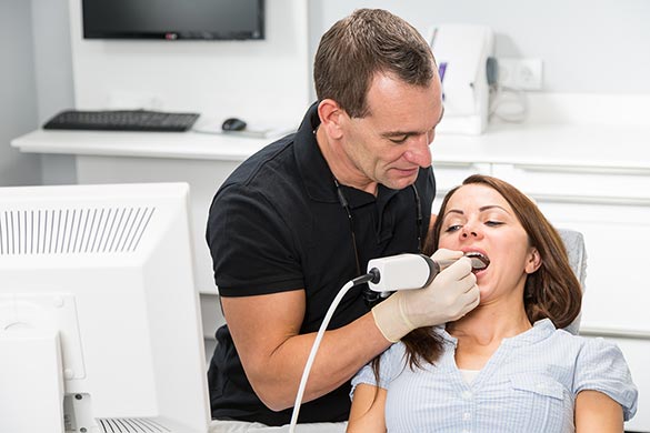
It is a state-of-the-art tool, manageable, and consisting of a small handheld device that is inserted into the mouth as if it were a normal dental instrument. The handheld device is easy to maintain and intuitive to use. In this way, the dentist can focus more on the patient and the maneuvers being performed.
INTRAORAL SCANNER VS TRADITIONAL IMPRESSION
Traditional impression materials, due to their characteristics and limited usage intervals, leave little room for maneuver. This means that decisive impression-taking maneuvers must be performed quickly. Moreover, in traditional impressions, in case of errors or failure to capture important details, it is necessary to redo the entire impression. All this, without taking into account the considerable discomfort for the patient during traditional impression-taking maneuvers.
There are several intraoral scanners on the market, but few are the leading companies in this sector. The dentists at our clinic have chosen to invest in one of the most performance-driven brands.
LATEST GENERATION INTRAORAL SCANNERS
The advantages in using this type of scanner are more evident when new and state-of-the-art instrumentation is used. This is what the professionals at our dental clinic in Milan do. The most advanced scanners, in fact, allow for the detection of dental optical impressions without the need to resort to opacification of oral surfaces. The latter requires the use of sprays and powders and more intervention by the dentist, resulting in greater discomfort for the patient.
Moreover, the instrument used must have a good scanning speed and must not stop during the examination. If it is necessary to take impressions of the second and third molars in particular, the dimensions of the scanner tip used are fundamental. In fact, they should preferably be small.
HOW IS THE EXAMINATION PERFORMED WITH THE INTRAORAL SCANNER?
Many dental clinics are still hesitant about introducing this scanner into their daily activities. They fear that its use requires complex specific training and think they are not yet ready.
Indeed, it is necessary to dedicate an initial period to learning how to best use the new equipment with training courses. In particular, operators who are already familiar with the digital world are advantaged. The staff at our dental clinic is always attentive to staying updated and has enthusiastically welcomed this innovation.
MAIN STEPS OF INTRAORAL SCANNING OF THE ORAL CAVITY
The examination through an intraoral scanner is very simple and quick – scanning the entire mouth takes about 5 minutes – and consists of these steps:
- The scanner tip is inserted into the oral cavity;
- The instrument is moved over and around the teeth;
- In case of interruption, the scanning can be resumed from where it left off;
- Chewing is detected by having the patient close their teeth with the scanner still inside the mouth;
- If there is a need to design dental appliances for correcting crooked teeth or various prosthetics, the patient can be involved. The patient can observe the dedicated screen where the obtained images are projected.
INTRAORAL SCANNER: THE FUTURE OF DIAGNOSIS IN DENTISTRY
Studio Motta Jones, Rossi & Associates is always attentive to providing its patients with innovative technologies, capable of ensuring greater comfort. We always carefully evaluate the future prospects for the use of each new tool. The intraoral scanner, today, can be used with excellent results in the design and production of prostheses and for implant surgery.
Furthermore, we achieve remarkable results using optical technology in the study of the mouth, even in the field of orthodontics. It is possible to study and simulate the treatment and the final result of orthodontic care. The information obtained from a facial scanner can also be combined with that provided by the intraoral scanner and even with CT scan data. This results in a “virtual model” useful in many dental therapies.


