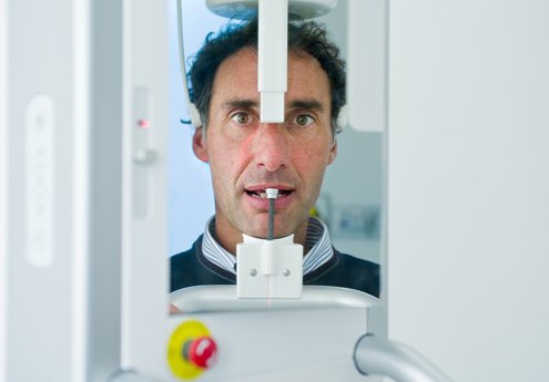
THE DIAGNOSTIC PRECISION OF DIGITAL DENTAL RADIOLOGY
Our dental clinic in Milan uses digital dental radiology systems. Radiographic examination serves to make diagnoses, evaluate, select, and recommend therapeutic treatments, as well as to conduct follow-up checks over time.
To identify the pathology affecting the patient and thus obtain a precise picture of the clinical situation, excellent image quality is necessary in diagnostic procedures. Digital dental radiology is an excellent diagnostic technique capable of ensuring the accuracy of the information to be collected. It is a diagnostic method that uses sophisticated hardware and software. These allow the detection, processing, and storage of radiographic images in the form of computer data via a computer. This radiology technique, introduced now for more than a decade, has marked a significant step forward compared to traditional film-based radiology.
THE ADVANTAGES OF DIGITAL DENTAL RADIOLOGY
Digital radiology represents a significant revolution in the field of dentistry and brings advantages for both the patient and the dentist. The advantages for the patient are:
- Greater speed in performing diagnostic examinations, thanks to the digital sensor that acquires and makes the images visible in a very short time;
- Less invasiveness because the exposure to X-rays required is significantly lower.
The advantages for the dentist are related to the quality of the images:
- Digital images undergo no alteration and remain unchanged over time, allowing consultation of the information even years later;
- Every single detail of the images can be modified to ensure greater readability, improving, for example, the contrast or brightness. Specific areas of the oral cavity that need further investigation can be enlarged.
DIFFERENT TYPES OF DIGITAL DENTAL RADIOLOGY
At our dental clinic in Milan, we perform various types of digital dental radiology:
- Digital intraoral radiology, aimed at analyzing specific dental elements to visualize the anatomy of the individual tooth components (crown, root, gums);
- Dental panoramic radiography – also called panoramic radiography, orthopantomography, or OPG. It provides a comprehensive frontal view of the dental anatomy of the patient’s skull in a single digital image. Teeth, mandibular and maxillary bone, maxillary and paranasal sinuses, temporomandibular joints, and gums are visible;
- Cephalometric radiography, which allows for a detailed frontal or lateral view of dental structures and the skeletal bases supporting the teeth. This allows for a specific analysis of the relationships between the teeth and the skull. The dentist is thus able to understand if there are any problems with dental malocclusion.


